
Inflammation
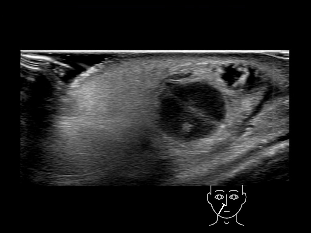
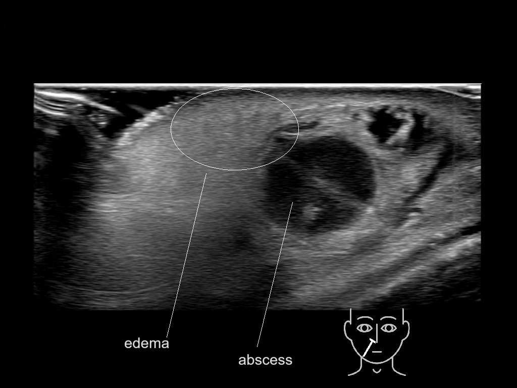
With ultrasound signs of inflammation can be visualized. Edema can be seen as a hyperechoic appearance of the subcutaneous fat, sometimes separated by hypoechoic fluid filled area’s, known as cobblestone appearance. Increased vascularization (hypervascularity) can be seen on colour Doppler. An abscess will appear as a fluid collection appearing as an irregular hypoechoic area with heterogeneous internal echoes and a thickened wall. Posterior acoustic enhancement can be present, and there is vascularity around but not within the mass. Under ultrasound guidance, abscesses can be managed by needle aspirations (18G) under antibiotic cover.
Study the first image to recognize the different layers. If you are sure about the layers, swipe to the second image to view the answer (if applicable).
Library
Hover over an image to view the secondary image or click on the image title for more information.